Description
Model:DW-T8 V4.0
Brand: Advanced Health Canada
Country of origin: Canada
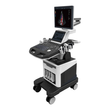
Real-Time 4D Color Doppler Ultrasound Scanner Machine DW-T8 V4.0
DW-T8 4D Color Doppler Ultrasound Scanner Machine is configured with a new imaging engine ultrasound scanner, which can significantly optimize imaging performance. It is a comprehensive imaging system engineered to meet today’s most demanding needs, from deep abdominal, vascular to superficial small parts.
This 4D color doppler ultrasound machine is equipped with Windows operating system DICOM3.0, widely applied to the abdomen, obstetrics, gynecology, heart, urinary system, small organs, superficial, blood vessels, pediatrics, newborns, musculoskeletal with its high-technology Auto-Adaptive Imaging Processing.
Specification:
-Stereo audio system
-Newly designed ergonomic console: The fully height-adjustable control panel enables optional positioning with just one touch
-Front and rear handles: Handles on both front and rear make T8 easy to be transported
-4 Active transducers ports: Improve your flexibility in moving through a wide range of applications
-4G RAM,120G SSD+500G HDD
-4 Wheels with locks: Highly maneuverable, highly mobile four-wheeled platform makes portable exams easy
- Main specifications and system of Troller 4D Color Doppler ultrasound
| 1.1 | Trolley type High-End Cardiac and Real-Time 4D Color Doppler Ultrasound |
| Probes | Convex probe Tran-vaginal probe Linear probe Micro-convex probe Cardiac probe 4D Volume probe |
| Applications and reports | Abdominal, OB/GYN, Cardiac, Urinary, Small Parts, Superficial, Vascular, Pediatrics, Advanced measurement software packages, report software packages, case management software packages, etc. |
| Performance | Carotid artery intima measurement thickness(IMT) Automatic spectral envelope measurement Full digital transmission and reception of beam synthesizer Color Doppler imaging(C) Pulse-Doppler Imaging(PW) Coherent Contrast imaging(CCI) Continuous-wave Doppler imaging(CW) B/C/D Real-time three synchronous imaging Power Doppler imaging(PDI) Direct power Doppler imaging(DPDI) M mode imaging Anatomic M mode imaging Color Doppler M mode imaging Elastography Tissue Doppler imaging(TDI) Strain rate imaging (SRI) Tissue harmonic imaging(THI) Fusion harmonic imaging(FHI) Speckle Reduce imaging(SRI) Panoramic imaging Deflection imaging Trapezoidal imaging Adaptive velocity optimization Freehand 3D Real-time 3D imaging(3D/4D) |
| Monitor | 21.5 inch HD LED, Multi-angle adjustable LED screen meets different clinic demands |
| Assistance monitor | 13.3-inch touch LED, It Brings doctors convenient operations and the time of examination is greatly shortened |
| Physical clipboard | Save the image on the left side of the screen, which can be directly saved or deleted. |
| PS | The system has the function of an on-the-spot upgrade |
| Presupposition | For different inspections of the viscera, preset the inspection conditions for the best image, reduce the adjustment of the operation, and the commonly used external adjustment and combination regulation. |
| Probe interface | 4 |
| Language | Chinese and English System, Chinese and English input, optional |
| Depth | ≥360mm; |
| PS | Extended imaging |
- Probe
| Convex probe | Fundamental Frequency:2.0MHz /2.3MHz /2.5MHz /3.0MHz /3.5MHz /4.0MHz /4.6MHz /5.0MHz /5.4MHz, Harmonic Frequency:4.0MHz /4.6MHz /5.0MHz, |
| Linear probe | Fundamental Frequency: 4.0MHz /4.6MHz /5.0MHz /6.0MHz /7.0MHz /8.0MHz /9.2MHz /10.0MHz /12.0MHz /13.3MHz, Harmonic Frequency:8.0MHz /9.2MHz /10.0MHz, |
| Transvaginal probe | Fundamental Frequency:3.0MHz /3.5MHz /4.0MHz /5.0MHz /5.4MHz /6.0MHz /7.0MHz /8.0MHz /10.0MHz, Harmonic Frequency:6.0MHz /7.0MHz /8.0MHz, |
| Micro-convex probe | Fundamental Frequency:3.0MHz /3.5MHz /4.0MHz /5.0MHz /5.4MHz /6.0MHz /7.0MHz /8.0MHz Harmonic Frequency:6.0MHz /7.0MHz /8.0MHz, |
| Cardiac probe | Fundamental Frequency:1.7MHz /1.9MHz /2.1MHz /2.5MHz /3.0MHz /3.4MHz /3.8MHz /4.2MHz /5.0MHz, Harmonic Frequency:3.4MHz /3.8MHz /4.2MHz, |
| 4D Volume probe | Fundamental Frequency:2.0MHz /2.5MHz /3.0MHz /3.3MHz /3.7MHz /4.0MHz /5.0MHz /6.0MHz, Harmonic Frequency:4.0MHz /5.0MHz /6.0MHz, |
3.2D Imaging Mode
| Gain | 0-100, Step 2 adjustable |
| TGC | 8 segment adjustable |
| Maximum focus point | ≥7, which can be moved throughout the whole process. |
| Speckle reduction | 0-5,5 level |
| Space Synthesis | 0-2,2 level(Liner probe: 3 levels, cardiac probe:0) |
| Dynamic | 30-180,35 level, step 5 adjustable |
| Line density | low、middle、high,3 level |
| Frame correlation | 0-4,4 level |
| Noise reduction | 0-5,5 level |
| Edge Enhancement | 0-5,5 level |
| Sound power | 2-10, 9 level |
| Greyscale | 0-67, 67 level |
| False-color | 0-67,67 level |
| Image style | Soft-Comparison,2 level |
| PS | The screen has a real-time display of voice power, probe frequency, dynamic range, pseudocolor, grayscale, and other 11 parameters that can be adjusted |
- Color Doppler Imaging Mode
| Blood gain | 0-100, Step 2 |
| Parameter display | Velocity、Variance |
| B-Restrain(B/W restrain) | 0-7, 7 level |
| Speed Through | 0-8, 8 level |
| Sampling number | 6-24, 7 level |
| Blood flow preferred | 0-8, 8 level |
| Filtering | 1-6, 6 level |
| Sound power | 2-6, 4 level |
| Noise reduction | 0-4, 4 level |


